



Superficial Radiotherapy for Skin Cancer: Why Positioning Matters
2026-07-15 18:07Why Small Skin Lesions Still Need Precise Positioning in Superficial Radiotherapy
The lesion may be small. The setup problem is not.
Consider a small lesion beside the nose.
The visible area is limited. The treatment head can reach it. The patient only needs to remain still for a short time.
Then the applicator approaches the skin.
The nose curves away from the beam. One side of the treatment area sits differently from the other. A nostril may require shielding. The patient turns their head slightly to breathe more comfortably.
The lesion still looks centered.https://www.kernelmedint.com/product/superficial-radiationidomestic-substitution-radiotherapy-for-skin-diseases
The setup may no longer be the same.
Nothing about that change appears dramatic. That is precisely why it matters.
In superficial radiotherapy for skin cancer, positioning is not simply about bringing the treatment head close to a lesion. It is about reproducing the planned relationship between the treatment field, the skin surface, the patient and any protective shielding.
A Small Lesion Is Not a Small Clinical Decision
Superficial radiotherapy is used for selected cancers on or close to the skin surface. Treatment decisions depend on factors such as lesion size, depth and anatomical location. Planning may involve skin markings, measurements, photographs and tracings so that the treatment area and patient position can be reproduced at later sessions.
This creates an important distinction:
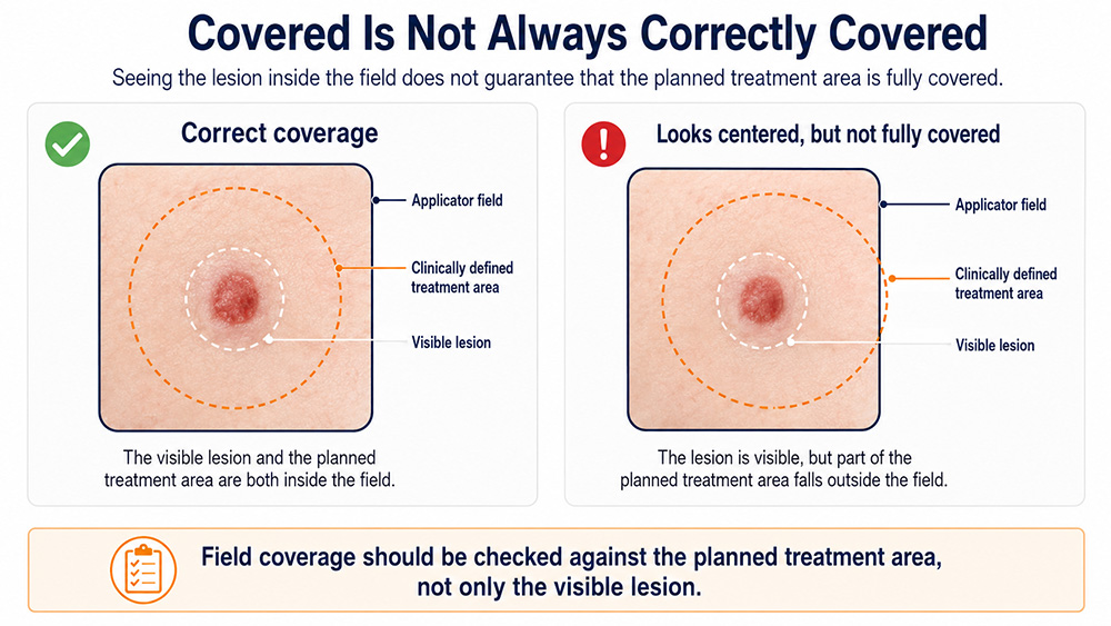
The visible lesion may fit inside the applicator while the planned treatment area does not.
The area that needs treatment is defined by the clinical team. It cannot be determined by looking only at whether the visible lesion appears centered beneath the treatment head.
A setup may look acceptable while still raising practical questions:
Does the field cover the complete planned area?
Is the treatment head approaching at the intended angle?
Has part of the skin curved away from the applicator?
Has the patient moved since the field was checked?
Is the shielding still in its planned position?
These are setup questions, not cosmetic details.
‘Covered’ Is Not Always ‘Correctly Covered‘
Seeing the lesion inside the field can create a reassuring impression.
The treatment head is in place.
The lesion is visible.
The field appears centered.
But visual centering alone does not confirm that the intended treatment geometry has been reproduced.
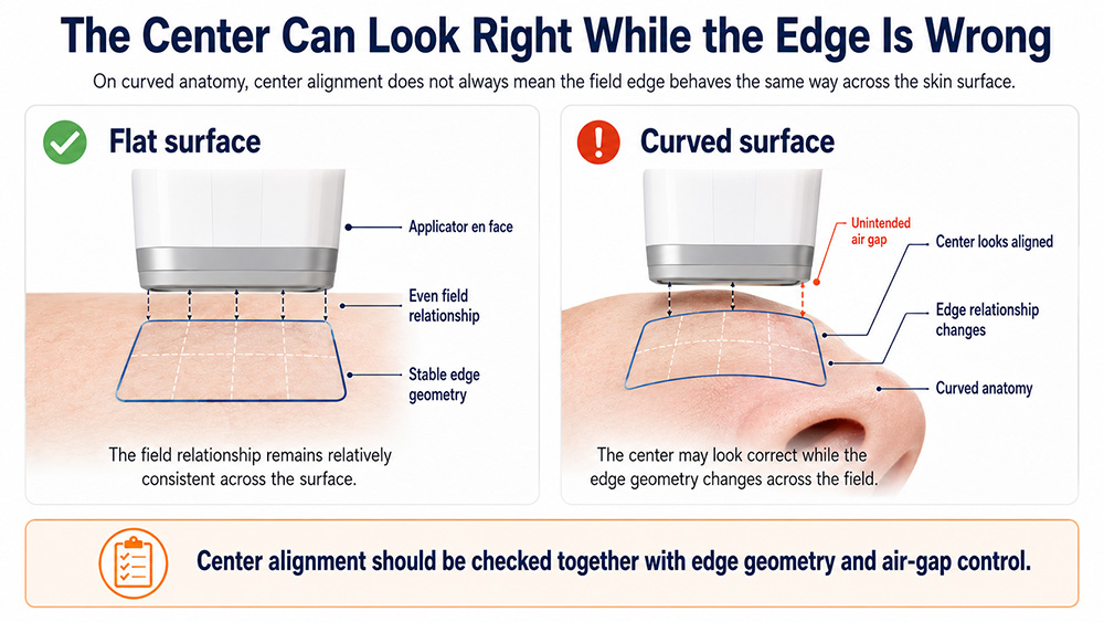
A published clinical implementation report for superficial radiation therapy described maneuvering the applicator so that the X-ray beam was en face to the treatment surface while minimizing the air gap between the skin and applicator. The report treated these steps as part of patient setup rather than optional refinements.
This is a better way to understand precise positioning.
It is not about moving the treatment head to approximately the right location. It is about checking whether the relationship established during planning is still present when treatment begins.
Curved Anatomy Changes the Question
A flat skin surface is relatively easy to visualize.
The applicator approaches the area, and the distance and angle remain reasonably consistent across the field.
The nose, ear, scalp and jawline are different.
The center of the field may look well positioned while one edge curves away. A small change in head angle can also change how the treatment surface faces the applicator.
Research on kilovoltage radiotherapy planning has identified irregular surfaces—particularly in the head and neck—as a limitation of simplified calculations based on a single average source-to-surface distance. Custom lead cutouts and shields add further geometric complexity.
This does not mean every curved surface requires the same correction.
It means the operator must look beyond the center of the field and follow the commissioned technique, treatment plan and medical physics procedures used by the facility.
The Nose Shows Why Shielding Is Part of the Setup
The nose combines several challenges in a very small space:
curved anatomy;
limited applicator access;
nearby sensitive structures;
changes caused by head rotation;
possible internal or external shielding.
Cancer Research UK describes how a narrow lead strip may be individually made to fit inside a nostril when a nasal skin cancer is treated. Moulds may also be produced to fit the treatment area and protect nearby tissue.
This makes shielding more than something added after positioning is complete.
The shielding is part of the geometry.
If it moves, it may no longer protect the intended area. If it overlaps the field, it may affect the planned arrangement. If it changes how closely the applicator can approach the skin, the setup needs to be checked again.
A small lesion can therefore require coordination between the treatment field, patient posture, applicator and shield—all within a few centimeters.
Repeat the Patient Position, Not Only the Machine Position
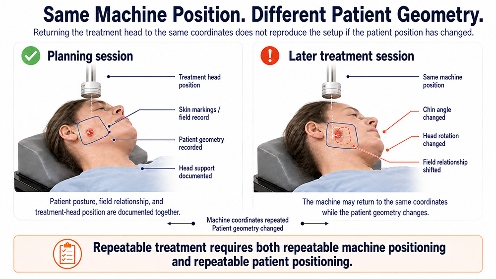
A treatment head can return to the same mechanical position while the patient does not.
The patient may lie slightly higher on the couch. Their chin may be lower. A shoulder may be raised. An arm may rest differently.
The machine coordinates can be repeated while the relationship between the machine and anatomy has changed.
That is why treatment teams may use photographs, measurements, tracings, skin markings, moulds and positioning supports. Cancer Research UK notes that radiographers use these references to help reproduce the correct position at each treatment session.
Consider a lesion near the ear.
At planning, the patient’s head is supported in a slightly rotated position. The field and shielding are documented in a photograph.
At a later session, the treatment head returns to its recorded position. Everything initially appears correct. When the operator checks the photograph, however, the patient’s chin is lower and the ear has rotated relative to the field edge.
The machine may not be the first thing that needs moving.
The patient position needs correcting.
Repeatable treatment requires both repeatable machine positioning and repeatable patient positioning.
An Air Gap Is Part of the Treatment Geometry
On a curved surface, part of the applicator may sit differently from the rest of the treatment area.
The lesion may remain visible, and the treatment head may feel stable. But an unintended air gap means the actual arrangement differs from the planned or commissioned setup.
Clinical implementation literature therefore describes minimizing the gap between the applicator and skin during superficial radiation therapy setup.
The correct response is not to invent a universal acceptable distance.
The operator should instead confirm that the geometry follows:
the treatment plan;
the applicator instructions;
the commissioned clinical technique;
the facility’s medical physics procedures.
This is especially relevant around the nose, ear, scalp, skin folds and other irregular surfaces.
What Robotic Positioning Can—and Cannot—Do
Robotic arm-assisted positioning can make it easier to approach different anatomical sites and adjust the treatment head without repeatedly moving heavy equipment or forcing the patient into an unstable position.
It may help the operator:
approach difficult facial or body locations;
make controlled treatment-head adjustments;
reduce repeated manual handling;
return toward a documented setup more efficiently.
The KernelMed XT-5601 Superficial X-Ray Radiation Therapy System combines robotic arm-assisted positioning with customizable beam limiters, stable radiation output and touchscreen control. These functions support the treatment workflow, but they should not be described as automatic clinical verification.
A robotic arm cannot determine:
the true clinical boundary of the lesion;
the required treatment margin;
the appropriate energy or dose;
whether shielding is correctly placed;
whether the patient has reproduced the planned posture;
whether the original setup was clinically correct.
A machine can reproduce a position.
It cannot decide whether that position should be reproduced.
A Practical Check Before Treatment Begins
A superficial radiation therapy setup should be reviewed as a sequence rather than a single glance at the lesion.
Before treatment, the team should confirm:
the patient and treatment site;
the patient position against available photographs, markings or measurements;
the planned field and treatment boundary;
the applicator or beam-limiting arrangement;
the angle, contact or distance required by the commissioned technique;
the position of any shielding;
whether the patient can maintain the posture.
The exact workflow will vary by institution, system and treatment plan. It should be defined by the responsible radiation oncology and medical physics team.
The important point is simple:
“The treatment head has arrived” is not the same as “the setup has been verified.”
What Clinics and Distributors Should Focus On
When evaluating a superficial X-ray therapy system, clinics should look beyond voltage range and positioning claims.
They should consider whether the treatment head can approach facial, scalp and limb lesions without forcing unstable patient positions. They should examine how easily the operator can see the treatment field, position shielding and make controlled adjustments.
Commissioning, quality assurance, safety controls, documentation, training and service support remain equally important.
Distributors should also avoid saying:
The robotic arm makes treatment accurate.
A more credible explanation is:
Robotic arm-assisted positioning helps clinicians approach different anatomical sites and adjust the treatment head with less manual handling. Treatment planning, patient positioning, field verification, shielding and final approval remain professional clinical responsibilities.
That statement gives the buyer a genuine reason to value the feature without suggesting that the equipment replaces radiation oncology expertise.
Conclusion: The Lesion Is Small. The Geometry Still Matters.
Small superficial lesions can look deceptively simple.
The visible field is limited. The equipment is close to the skin. Treatment may last only a few minutes.
Yet the setup can still involve curved anatomy, a clinically defined treatment area, applicator alignment, air-gap control, individualized shielding and repeated patient positioning.
Robotic positioning can make this workflow more practical.
Photographs, markings, measurements and supports can make it more reproducible.
But no single device feature replaces the central requirement:
The correct treatment geometry must first be planned, reproduced and verified.
The lesion may be small.
The responsibility is not.
FAQ
Why does positioning matter for a very small skin lesion?
The planned treatment area may extend beyond the visible lesion. A small field can leave less room for changes in patient posture, applicator position or shielding.
Why are the nose and ear difficult superficial radiotherapy sites?
These areas have curved surfaces, limited working space and nearby structures that may require protection. Small changes in patient position can also alter how the treatment surface faces the applicator.
Should the applicator be placed directly against the skin?
The required relationship depends on the applicator, commissioned technique and treatment plan. Published superficial radiation therapy workflows describe en face positioning and minimizing unintended air gaps.
How is the setup reproduced at later treatments?
Treatment teams may use photographs, measurements, tracings, skin markings, moulds and positioning supports to reproduce the planned patient and treatment geometry.
Does robotic positioning guarantee the correct setup?
No. It assists treatment-head adjustment. Clinical staff must still confirm the patient position, treatment field, applicator arrangement and shielding.
What should clinics evaluate when choosing a superficial X-ray system?
Clinics should evaluate positioning range, field-shaping options, treatment controls, output stability, safety systems, commissioning requirements, quality assurance, training and technical support.
References
1.Cancer Research UK. Superficial Radiotherapy to the Skin. Planning, treatment positioning, photographs, measurements, tracings, moulds and patient-specific shielding.
2.Lee YC, Davis SD, Romaguera W, et al. Implementation of Superficial Radiation Therapy Using SRT-100 Vision for Non-Melanoma Skin Cancer in a Radiation Oncology Clinic. Journal of Applied Clinical Medical Physics. 2023;24(6):e13926.
3.Nikandrovs M, McClean B, Shields L, et al. Clinical Treatment Planning for Kilovoltage Radiotherapy Using EGSnrc and Python. Journal of Applied Clinical Medical Physics. 2023;24(2):e13832.
4.Furstoss C, Dunscombe P, Arsenault C, et al. CPQR Technical Quality Control Guidelines for Kilovoltage X-Ray Radiotherapy Machines. Journal of Applied Clinical Medical Physics. 2018.
5.Kernel Medical Equipment Co., Ltd. XT-5601 Superficial X-Ray Radiation Therapy System for Skin Lesions.