



Reflectance Confocal Microscopy in Skin Cancer Workflows: When Dermoscopy Is Not Enough
2026-05-27 15:13Reflectance Confocal Microscopy in Skin Cancer Workflows: When Dermoscopy Is Not Enough
In many dermatology settings, dermoscopy is the first imaging step after routine visual examination. It improves lesion assessment, supports triage, and helps clinicians decide which lesions are reassuring, which need monitoring, and which may require biopsy. But experienced clinicians also know that not every lesion becomes clear under dermoscopy. Some pigmented lesions remain equivocal. Some facial lesions are difficult to assess with confidence. Some basal cell carcinoma cases raise questions about margins, residual disease, or whether another biopsy is immediately necessary.
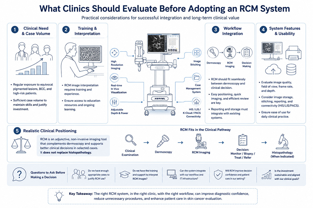
This is where reflectance confocal microscopy (RCM) becomes relevant. RCM should not be described as a replacement for dermoscopy, and it should not be presented as a substitute for histopathology. Its value is more practical than that. In selected cases, it adds a higher level of non-invasive imaging between dermoscopy and biopsy, helping clinics refine management decisions when surface-level assessment is not enough.
For clinics and distributors evaluating a reflectance confocal microscopy system, the real question is not whether RCM sounds advanced. The better question is where it fits in daily lesion management, which patients and lesion types justify its use, and what system capabilities matter in real clinical workflows.
Dermoscopy Is the Starting Point, Not the Whole Answer
A sensible skin cancer workflow does not begin with RCM. It begins with clinical examination and dermoscopy. Dermoscopy remains a practical front-line tool because it is fast, widely used, and highly valuable for routine lesion screening. In other words, the role of RCM only makes sense when its place is defined relative to dermoscopy, not in opposition to it.
The limitation is that dermoscopy is still a surface-oriented method. It can improve pattern recognition, but it does not always resolve uncertainty in lesions that are clinically suspicious, dermoscopically equivocal, located in cosmetically sensitive areas, or difficult to interpret because of background sun damage or atypical morphology. In these situations, moving directly from dermoscopy to biopsy may be appropriate in some cases, but not every uncertain lesion benefits from immediate excision as the first response.
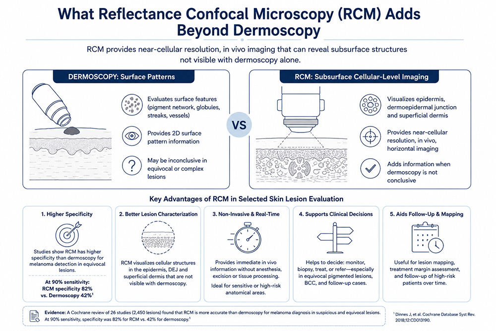
This is why RCM has become important in selected lesion workflows. It offers in vivo imaging at near-cellular resolution and can help clinicians examine structures in the epidermis, dermoepidermal junction, and superficial dermis more closely. That extra level of detail is what makes it useful as an adjunctive imaging step rather than just another visual device.
What RCM Adds Beyond Dermoscopy
The value of RCM is not simply that it produces sharper images. Its clinical relevance comes from the type of information it adds. Dermoscopy helps clinicians assess surface patterns. RCM goes further by enabling horizontal, real-time imaging of superficial skin architecture at a much higher level of detail.
This matters most when lesion management decisions are uncertain. A Cochrane review on RCM for melanoma diagnosis found that RCM was more accurate than dermoscopy in studies involving lesions suspicious for melanoma and in more difficult, equivocal lesion populations. In a model assuming a fixed sensitivity of 90% for both tests, specificity was 82% for RCM versus 42% for dermoscopy in lesions suspicious for melanoma. That finding is clinically relevant because improved specificity can translate into fewer unnecessary excisions of benign lesions without abandoning diagnostic caution.
That does not mean RCM should be marketed as a “biopsy avoidance machine.” It means RCM can improve confidence in selected workflows where dermoscopy alone leaves uncertainty. In real practice, that may help refine management decisions, support lesion selection for biopsy, or justify closer follow-up in carefully chosen cases.
Where RCM Is Especially Useful in Skin Cancer Workflows
1. Equivocal Pigmented Lesions
One of the most established roles of RCM is in the evaluation of equivocal pigmented lesions. These are lesions that are not clearly benign, but not convincingly malignant on dermoscopy alone. This is exactly the kind of situation where RCM adds value: not by replacing clinical judgment, but by providing another layer of information that may improve decision-making.
Clinically, this is important because equivocal lesions are common in dermatology practice, especially in patients with multiple atypical lesions, sun-damaged skin, or a history of skin cancer. In these cases, better specificity matters. The goal is not to “watch everything” or “excise everything,” but to make better-informed choices.
2. Facial or Cosmetically Sensitive Areas
Lesions on the face, ears, or other cosmetically important locations create a different clinical pressure. A low threshold for biopsy may still be appropriate, but clinicians often want more confidence before performing an invasive procedure in visible areas. RCM can be valuable here because it offers a non-invasive method to examine suspicious lesions more closely before moving forward.
This does not mean biopsy becomes unnecessary. It means the pathway becomes more thoughtful. For some lesions, RCM may support the decision to biopsy. For others, it may support more targeted monitoring or help clarify whether a lesion really deserves immediate excision.
3. Basal Cell Carcinoma Assessment
RCM is also relevant in basal cell carcinoma workflows. It has been used not only for diagnosis but also for lesion mapping and assessment of residual disease in selected settings. A prospective study by Navarrete-Dechent and colleagues showed that RCM could confirm residual basal cell carcinoma at clinically negative biopsy sites before Mohs surgery, highlighting its practical value in difficult management scenarios.
This is a more useful way to discuss RCM in BCC than simply saying “RCM can diagnose BCC.” The real point is that it may support decision-making where surface examination is not enough, especially when clinicians are trying to understand whether residual tumor is still likely or whether lesion extent remains unclear.
4. Follow-Up of High-Risk or Previously Assessed Lesions
RCM can also be helpful in the follow-up of selected lesions over time. In practices that monitor high-risk patients or lesions that do not yet meet a clear threshold for excision, RCM may contribute non-invasive longitudinal assessment. That is especially relevant in specialized dermatology centers with a structured lesion-monitoring workflow.
Again, the key phrase is “selected cases.” RCM is not necessary for every follow-up lesion, and not every clinic needs to build an advanced imaging pathway. But for centers already working with higher-risk lesion populations, it can support more informed follow-up decisions.For a broader overview of non-invasive skin cancer imaging, see KernelMed's article on RCM for skin cancer diagnosis.
What Clinics Should Evaluate Before Adopting a RCM System
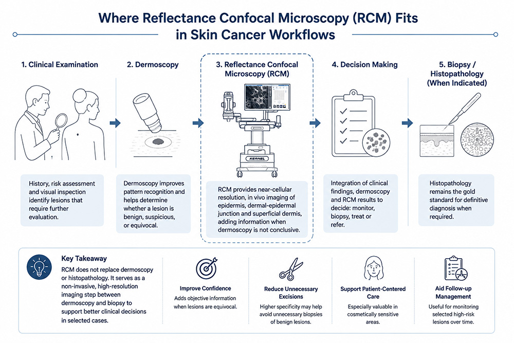
A common mistake is to think that if the clinical rationale for RCM imaging systems is sound, the equipment decision is automatically simple. It is not. Clinics should evaluate not only whether RCM is useful in principle, but whether a specific system can be integrated into everyday clinical work without adding unnecessary complexity.
The first thing to evaluate is case mix and lesion volume. A clinic with regular exposure to equivocal pigmented lesions, facial lesions, high-risk patients, or skin cancer workups is far more likely to benefit from RCM than a clinic with very limited diagnostic imaging needs.
The second point is training and interpretation. RCM does not create value on its own. Its usefulness depends on whether clinicians can acquire interpretable images and incorporate them into decision-making. This is why training and team readiness matter just as much as hardware specifications.
The third point is workflow integration. An RCM system should fit the way a clinic already works. Can it be used efficiently after dermoscopy? Can images be reviewed without slowing down the entire visit? Can reports be stored, retrieved, and incorporated into the patient record without creating a data bottleneck?
The fourth point is image acquisition capability. Clinics should look at whether the system can consistently produce usable images, whether lesion positioning is manageable, and whether the platform supports practical examination of different sites.
This is where product-level details become meaningful. For example, KernelMed’s current RCM platform specifications include an 830 ± 5 nm laser, a 40× objective with NA 0.8, central optical resolution below 1.25 μm, central depth of field below 5.0 μm, a 500 μm × 500 μm scan field, 1024 × 1024 image resolution, and a frame rate of at least 15 fps. From a clinical workflow perspective, these are not just engineering numbers. They matter because they influence image clarity, examination usability, and how efficiently the system can be used in daily practice.
Additional workflow-related features are equally important. The specification sheet also lists adjustable laser power, manual positioning adjustment, imaging depth adjustment, image stitching, image storage, and a built-in case management system with HIS, LIS, K-cloud, and PACS connectivity, along with integrated report printing. These details are directly relevant to adoption because they determine whether the RCM platform can function as part of a real documentation and reporting workflow rather than as a stand-alone imaging device.
The fifth point is realistic clinical positioning. An RCM system should be adopted because the clinic needs an imaging step beyond routine visual assessment and dermoscopy in selected cases, not because it wants a premium device for marketing.
The strongest clinical position for RCM is as an adjunctive, non-invasive imaging tool that improves assessment quality in the right workflows.
What This Means for Distributors
RCM is not an entry-level dermatology device, and distributors should not position it like one. It is also not a product that should be sold with exaggerated promises such as “non-invasive replacement for biopsy.” That is the wrong message and it weakens credibility.
A more appropriate positioning is that RCM is a high-value adjunctive imaging tool for clinics that already manage difficult lesion assessment, equivocal pigmented lesions, skin cancer screening, or complex follow-up workflows. The best-fit customers are usually dermatology departments, skin cancer clinics, specialized lesion centers, and advanced dermatology practices that already use dermoscopy and are looking for a higher-resolution imaging layer.
Distributors should also understand that the discussion cannot stop at clinical indications. Buyers will ask practical questions: Is the system easy to move? Can it be used in a normal examination room? How are images stored? Can the system connect to existing hospital information workflows? Does it support reporting? Is the arm or positioning system practical for different body sites?
This is exactly why system-level features matter. In KernelMed’s platform, the trolley-based structure, monitor-based operation, adjustable cantilever support, image stitching, case management, and hospital data connectivity are not small details. They shape how the product will actually be experienced in a clinic environment.
The right distributor message is therefore not “RCM is sophisticated.” The stronger message is: this is a non-invasive imaging platform for clinics that need more than routine visual assessment and dermoscopy, and it should be evaluated as part of a complete lesion-management workflow.
RCM Has a Defined Role, Not an Unlimited One
One reason RCM is easy to overstate is that it sits in an attractive middle ground: more detailed than dermoscopy, less invasive than biopsy. But this middle position is exactly why it must be described carefully.
RCM does not replace histopathology. It does not solve every diagnostic problem. It does not remove the need for clinical judgment. Its imaging depth is limited to superficial skin structures, and interpretation still depends on expertise.
Yet when used in the right setting, it can be genuinely valuable. It can improve confidence in selected equivocal lesions. It can help in cosmetically sensitive areas where clinicians want better pre-biopsy assessment. It can contribute to selected BCC and follow-up workflows. And it can help advanced dermatology centers build a more refined, non-invasive diagnostic pathway.
That is the right level of claim. It is credible, useful, and clinically relevant.
Conclusion
Reflectance confocal microscopy has a meaningful place in skin cancer workflows, but only when its role is understood correctly. It should not be presented as a replacement for dermoscopy, and it should not be marketed as a substitute for histopathology. Its real value lies in selected situations where clinicians need more information than dermoscopy can provide, but want to remain non-invasive before deciding on biopsy or treatment.
For clinics, the adoption decision should be based on lesion volume, case complexity, workflow integration, reporting needs, and image interpretation capability. For distributors, the key is to position RCM for the right customers and explain it as an adjunctive imaging system rather than a universal diagnostic shortcut.
In the right hands and the right workflow, RCM is not just another imaging device. It is a practical bridge between dermoscopy and biopsy.
FAQ
1. What is the role of reflectance confocal microscopy in skin cancer workflows?
RCM works as an adjunctive, non-invasive imaging step between dermoscopy and biopsy in selected cases. It is particularly useful when lesions remain equivocal after dermoscopy or when clinicians want more information before performing an invasive procedure.
2. Does RCM replace dermoscopy?
No. Dermoscopy remains the front-line imaging tool in most lesion assessments. RCM is best understood as a higher-resolution adjunct used when dermoscopy alone does not answer the clinical question clearly enough.
3. Can RCM replace biopsy or histopathology?
No. Histopathology remains the definitive diagnostic standard when tissue confirmation is required. RCM may help refine management decisions, but it does not replace pathology.
4. In which skin cancer scenarios can RCM be especially helpful?
RCM is especially useful in equivocal pigmented lesions, cosmetically sensitive areas such as the face, selected basal cell carcinoma workflows, and follow-up of certain high-risk lesions.
5. What should clinics evaluate before adopting an RCM system?
Clinics should evaluate lesion volume, case mix, staff training, image interpretation ability, workflow integration, image storage and reporting, and whether the system fits existing hospital information infrastructure.
6. What should distributors focus on when positioning an RCM system?
Distributors should focus on workflow fit, target customer profile, reporting capability, connectivity, image-management functions, and realistic clinical positioning rather than exaggerated claims.
References
Levine A, et al. Introduction to reflectance confocal microscopy and its use in clinical practice.
KernelMed product parameter sheet for current reflectance confocal microscopy platform